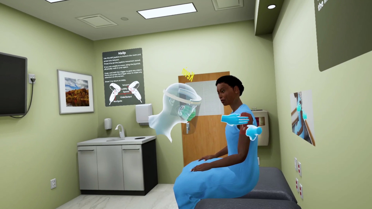
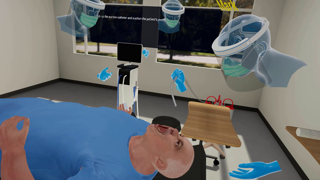
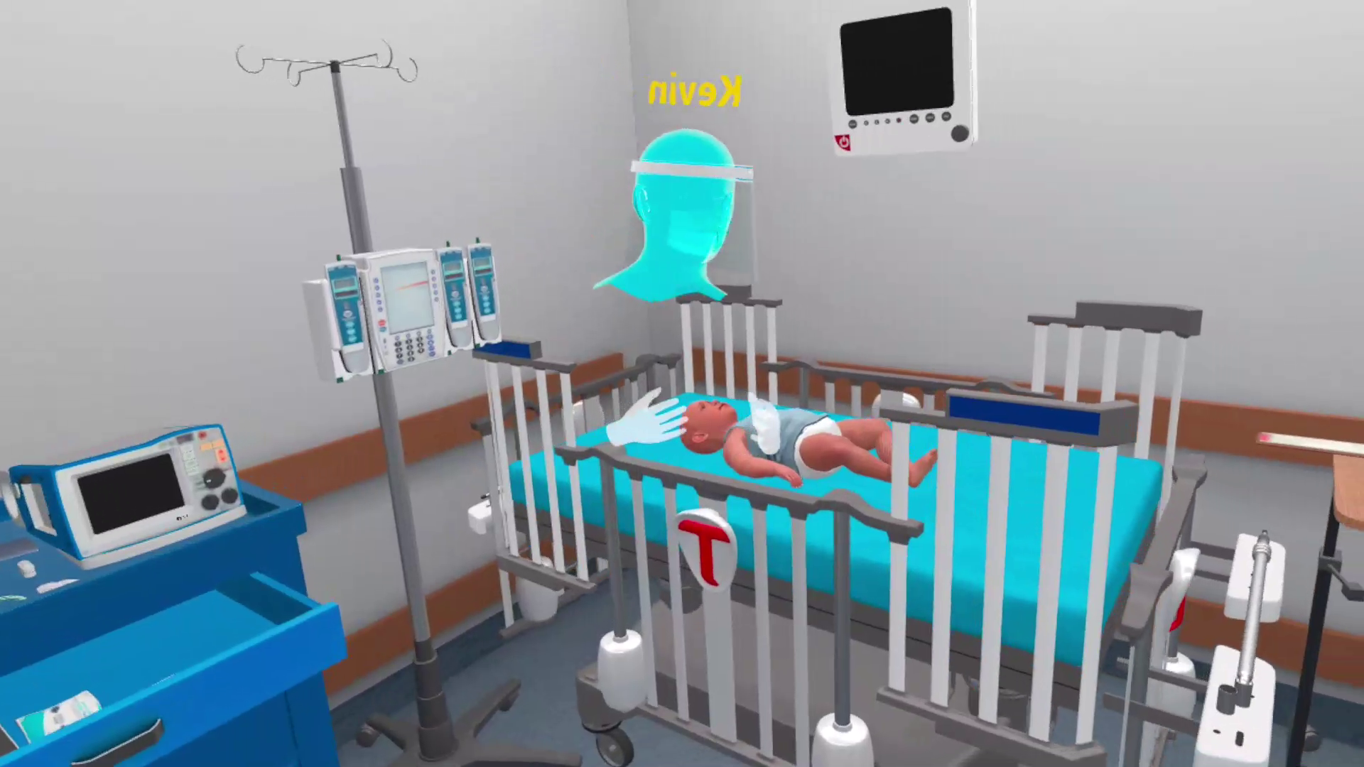
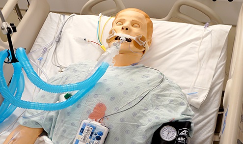
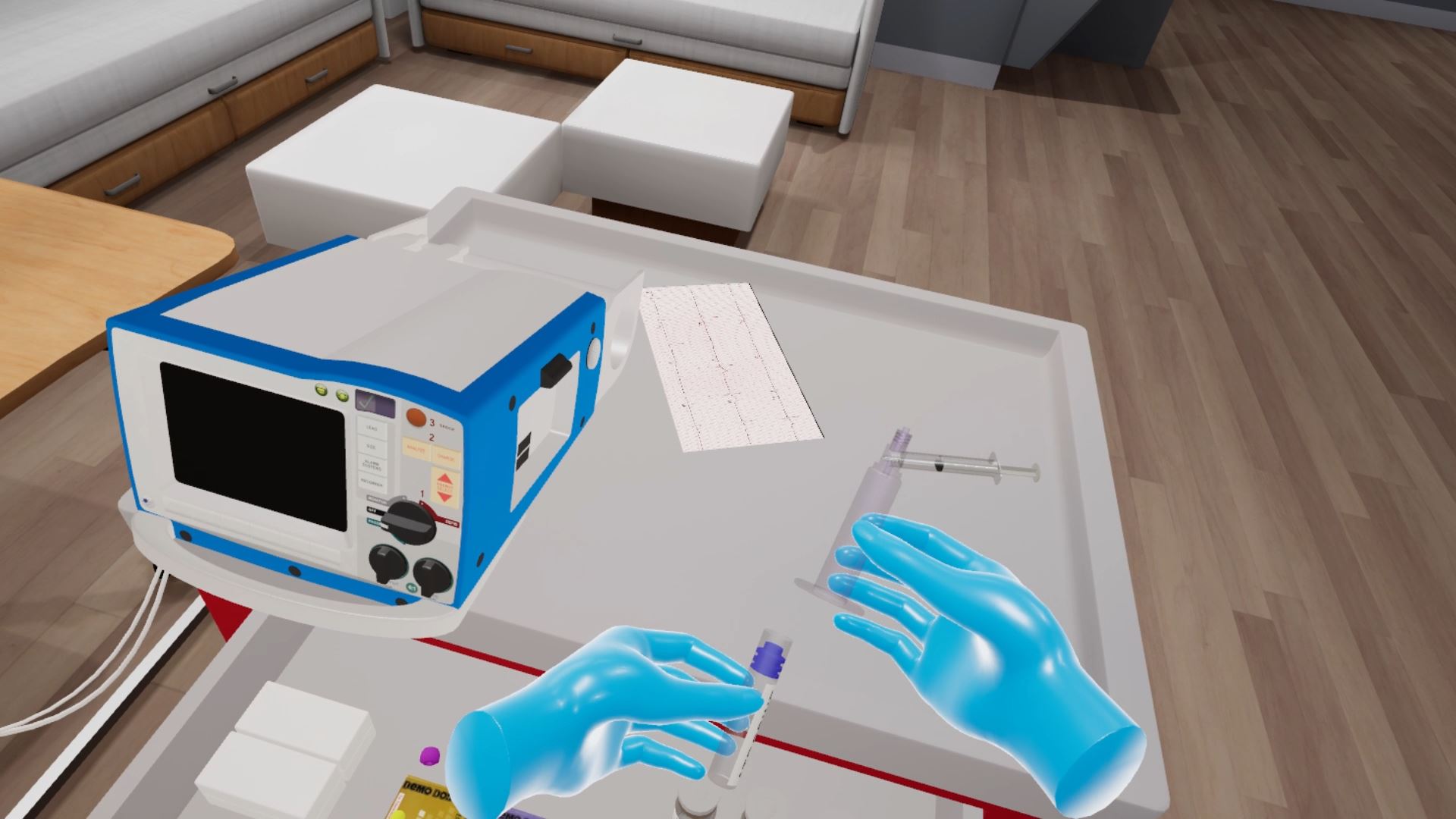
Interactive 3D models don’t just have applicability in product marketing. They’re finding a home in medical training, including visualization in both AR and VR formats. How is this coming together and how does it hold potential to transform anatomical education?
How Is XR Transforming Anatomical Education?
XR is transforming anatomical education because it provides 3D models — something many courses lack. According to one study, 69.8% of medical students’ top complaint when learning anatomy is the lack of visual resources. Naturally, seeing up-close and detailed examples is critical when studying something so complex.
Unlike cadavers, physical models or textbook images, 3D elements are interactive. Students can move, zoom in or rotate them to get a better view of anatomical structures, helping them better understand spatial relationships. It’s also been demonstrated that learning with 3D models has efficacy in boosting memory recall, compared with traditional methods and non-immersive benchmarks.
Interactive 3D models through immersive XR interfaces can also function as anatomical structures do during life or certain medical events. For example, a replica of a vascular system can show students what a heart attack looks like. They can see how various internal structures react to disease or trauma.
The Technological Advancements Making It Possible
XR-powered interactive 3D models only exist today because of a combination of multiple technological advancements. One of the most significant is headsets because they make hands-free viewing possible. Students probably wouldn’t experience the same benefits if they had to hold their phones for the entire lesson.
Spatial mapping is another significant technological advancement that makes interactive 3D anatomical models possible. It enables students to place an augmented element in the real world, seamlessly aligning it with their surroundings.
The entire concept of interactive 3D models hinges on interaction technology. It encompasses gestures, voice commands, touch and controllers. For medical students to be able to zoom in, rotate or move a 3D element, they need to be able to register their commands and respond accordingly.
5 Benefits of XR in Medical Training
Students and professors can benefit substantially from using 3D models for training. This can speed up medical processes, potentially accelerating students’ learning rates and improving their learning outcomes.
1. Interactive Real-World Visualization
Through XR – including AR experiences through devices like Magic Leap 2 – students can visualize complex anatomical structures in the real world. The 3D model appears directly in front of them without blocking their view of their professor’s gestures, their classmates’ expressions or the room they’re in. This way, they have an easier time transitioning from a learning stage to a practicing one.
2. Enhanced Accessibility for Remote Learners
Immersive 3D learning also improves distance education for students. Anyone taking online classes can benefit from this technology’s interactiveness. It can help them achieve the same exam scores and learning outcomes as their peers despite their different circumstances.
3. Accessibility When Cadavers Are Unavailable
A steady supply of cadavers isn’t guaranteed, as many learned during the COVID-19 pandemic. Many students had to rely on dated textbook images and physical models. Those with immersive 3D models could continue studying anatomical structures despite the shortage. In similar future scenarios, this technology could prevent a generation of students from falling behind.
4. Enhanced Learner Engagement Rates
As noted, immersive learning increases students’ retention rates by making them more interested and involved in their studies. It can cultivate better engagement by making lessons immersive and realistic. Instead of reading a textbook or struggling to interpret poorly scanned black-and-white textbook images, they get to physically interact with the material.
5. Better Access to Rare Anatomical Structures
Immersive learning through simulated 3D models can provide medical students with a broader variety of anatomical structures to help them establish a better baseline for what they should expect in practice. It can also give them better access to rare or unusual anatomy, improving their understanding of edge cases.
How XR Will Further Advance Medical Education
There is potential for further innovation in medical education as XR advances medical training and professionals explore new use cases. If professors and students continue using it, they’ll find new ways to benefit from the technology.
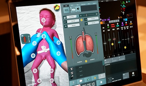
One likely outcome is that students begin using XR in real-time while practicing. For example, they can use it to view artificial vital signs, ultrasound images or X-rays to enhance their understanding and accelerate their learning.
Whatever happens, medical training is all but guaranteed to continue using XR since it offers such great learning benefits. One research team described AR as remarkable, noting it helped medical students better understand 3D anatomical structures and score well on exams.
The Future of Medical Training Involves AR
Immersive learning through AR and VR lets students view the world around them while studying and practicing, a critical factor when working with delicate internal structures. Considering it has already made its way into surgeries and therapy, there’s no doubt it will continue making waves in medical education.
Quelle:
